NeuroART Gallery
The NeuroART Gallery is a space where the members of the SENC can exhibit original collections about the nervous system. The SENC Board can also select interesting works and collections of authors who are not members of the Society.
The images can be sent to the email electrónica secretaria.tecnica@senc.es, as follows

TIFF, JPG OR PDF FORMAT

TITLE

AUTHOR OR AUTHORS NAME
Kathleen Sluka – Células de vida, Arte original
 Kathleen Sluka is a Professor at the University of Iowa focusing on the neurobiology of musculoskeletal pain and analgesia. In her spare time, she is a science artist with a focus on the cell biology in normal and disease states with an emphasis on neuroscience-related images. Her training as a physical therapist, anatomist, and neurobiologist serves as the inspiration and the scientific basis for her work.

H. Kathleen – Diseños
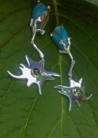 Kathleen is an artist, poet and jewelry designer who has specialized in creating highly personalized fine jewelry that tells her special story through the magic of metals and precious stones. She appreciates the creation of jewelry as a “trip” (with a vision she wants to express), instead of a “destination”, and she loves personalized attention, creative options, deepening in gems, and handmade art.
Lia Cook – Works
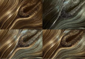
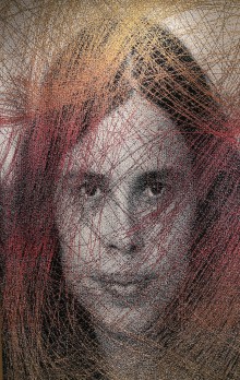 Her current practice explores the sensuality of the woven image and the embodied emotional connection to memories of touch and cloth. Visualization of data collected from behavioral studies conducted within an exhibition is woven back into the work. She has used DSI (Diffusion Spectrum Imaging of the brain) and TrackVis software to look at the structural neural connections between parts of the brain and integrate these “fiber tracts” with the actual fiber connections that make up the woven translation of a face.
Fernando de Castro – Work
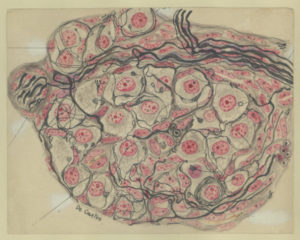 Fernando de Castro (Madrid, 1896-Madrid, 1967) was one of the last direct disciples of Santiago Ramón y Cajal and pre-eminent member of the Spanish Neurological School. His skills as a draftsman border on the highest level of the School itself, together with Río-Hortega. Fernando de Castro is universally known for his discovery of arterial chemoreceptors, which he located in the glomus caroticum (or carotid body). Here is an original drawing of him, published in one of his fundamental works in 1926, which shows the glomus cells that detect changes in the composition of the blood. This drawing served as an image for the SENC congress of 1995, held in Valladolid. This original drawing belongs to the Archivo Fernando de Castro (Censo-Guía de Archivos de España e Iberoamérica #ES.28079.AFC; Madrid, Spain), que en 2017 la UNESCO incluyó en the Memory of the World International Register of the Human Heritage, as `Archives of Santiago Ramón y Cajal and the Spanish Neurohistological School´
Legado Cajal
With this term, it is named the collection formed by a series of belongings of Santiago Ramón y Cajal, mostly scientific, that he wanted to be preserved in the Cajal Institute upon his death. It is composed of a valuable and varied material that, to quote succinctly, is made up of Furniture (work table, preparation cupboards …), Laboratory equipment (microscopes, microtomes, photo cameras …), Scientific material (histological drawings, anatomical oils, photos of microscopy, histological preparations, manuscripts and laboratory notebooks …), Awards and recognitions (medals, medals, diplomas …), Epistolary, Leisure material (pocket camera, photographic stereoscopic plates of his trips, photographs to color …), complete scientific library, objects of personal use (passport, identification card, glasses, cane …), etc.

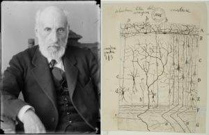 Original drawing of Cajal, representing a scheme of the afferent and efferent cortical projections. The efferents are determined by the projections, outside the cortex, of the axons of pyramidal cells of various sizes that, depending on the cortical stratum where they are located, will project to different subcortical structures. Note how some of these axons bifurcate in two when entering white matter, projecting one of the branches subcortically and the other continuing through the white substance to reach, in some cases, the contralateral hemisphere, via the corpus callosum. In other cases, the projection would remain ipsilateral, interrelating neighboring association cortical areas. The afferent projections to the cortex are represented, mainly, by robust axons from the thalamus, which enter and are mainly arborized in layer IV of the cerebral cortex. Other details shown in this drawing are different cell types of layer I, and a cell type of deep layers whose axon ascends to arborize layer I, known as Martinotti cells.
Michele Banks – Artologica
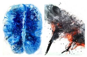
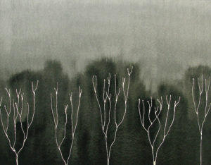 Michele Banks creates art – mostly paintings – exploring themes from science and medicine, including images such as viruses, bacteria, and plant and animal cells. She looks at how these organisms affect humans, and in turn how we affect them, through climate change, antibiotic use and other impacts on the earth. She finds watercolor to be an ideal medium for this type of work because it can be transparent or translucent, allowing her to hint at what is happening beneath the surface. Watercolor also naturally flows into fractal patterns such as those seen in the nervous and circulatory systems, as well as in tree branches and river systems – in fact, everywhere in nature.
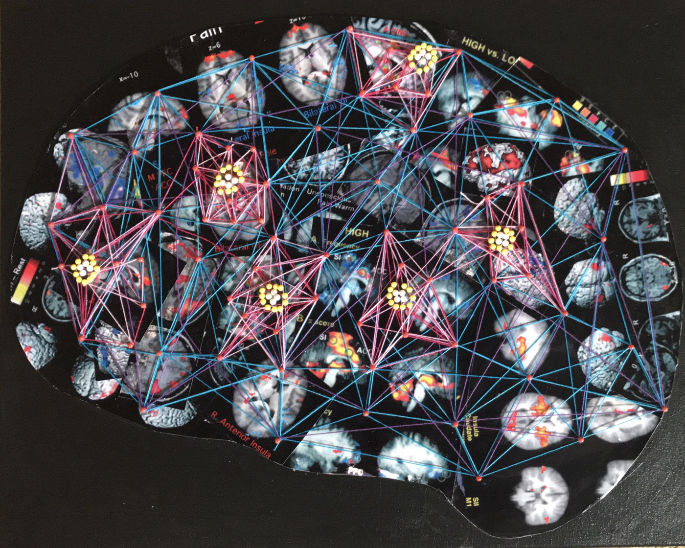
Laura Bundensen – Stitching together Art & Neuroscience
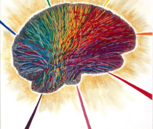 In her work, Laura Bundensen explores the fascinating terrain of the brain in texture and color. Her aim is to translate real neuroscience information into abstract fiber art that creates a sense of joy and wonder. Her work includes hand embroidery, vibrant color and a playful use of materials that represent the wonderful, magical human brain.
Lidó Rico – Genooarchitectures
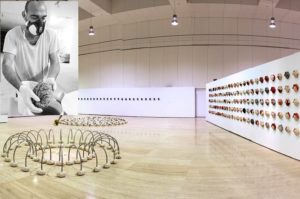 Genooarchitectures is the result of the interaction between the work of the sculptor Lidó Rico with the neurophysiologist Kuei Y.Tseng (Department of Anatomy and Cell Biology at the University of Illinois in Chicago) and the neurobiologist José Luis Ferran (Department of Human Anatomy and Psychobiology, of the Faculty of Medicine at the University of Murcia). From the mold of different brains, the work deals with the origin, the basic functions, the inadequate functioning and diverse cerebral pathologies; producing an approach between art and science leading to the complex being seen as something close and tangible. These Neurosculptures develop a discourse that establishes a new vision about our shared and fascinating machinery of thought, considering vitally important the active and social function that art should play as a vehicle of knowledge.
Luis Miguel Gutiérrez – From the Universe to the Brain
 Researcher at the Institute of Neurosciences (CSIC-Universidad Miguel Hernández, Alicante, Spain) and graphic artist, Luis Miguel Gutiérrez combines in his work Art and Science with the creation of a pictorial technique based on his experimentation with behavior as fluids of cytoskeletal cell structures. In recent years, this work explores the macro and microscopic worlds, which, beginning in the Big Bang, end with a recreation of the textures of the brain, as the natural structure that ends up giving the universe the property of self-consciousness. The exhibition recently exhibited at the Museum of Contemporary Art in Elche represents a unique way to put together Art, Science and dissemination. You can see his work in these links:
Elizabeth Jameson Portfolio
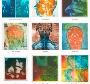
In 2013, Elizabeth Jameson, an American artist, makes some of her graphic works available to the SENC. Elizabeth creates prints and digital impressions based on MRI scans of the brain, including hers. Her work use intense colors and give the anatomical structures of the brain an dreamlike quality. Do not miss her creations:
Greg Dunn and Brian Edwards – Self Reflected
 Dr. Greg Dunn (artist and neuroscientist) and Dr. Brian Edwards (artist and applied physicist) created Self Reflected to decipher the nature of human consciousness, representing the relationship between macroscopic images of the brain in three dimensions and microscopic behavior of the neurons. Through a technique of reflected micro-schemas, they achieve images of great beauty. To learn more about the technique and about the work, visit their website:
NeuroART – MBF Bioscience – Gallery of images and contest
NeuroART is a contest sponsored by MBF Bioscience, a company dedicated to quantitative microscopy equipment for life sciences researchers. Images about the nervous system are shown from two perspectives:
 i) one is technical, through dyes and tracers to see individual neurons, neuronal circuits and brain structure, as well as functional magnetic resonance images.
ii) the other one is abstract, in which real images obtained in the microscope are modified with artistic techniques.
Be a member here
Contribute to the progress of neuroscience!

Collaborate with society
Do you want to publish your news or events in our schedule?