Portadas de revistas científicas
Uno de los objetivos de la SENC es la divulgación de imágenes científicas de miembros de la SENC que hayan sido objeto de publicación en Portadas de Revistas Científicas. Cualquier miembro de la SENC puede enviar las imágenes y/o composiciones publicadas como portadas en revistas científicas para su difusión en la página web de la SENC.
Las portadas de revistas científicas se enviarán a la dirección electrónica secretaria.tecnica@senc.es, del siguiente modo:

FORMATO JPG

NOMBRE DEL SOCIO QUE LA PRESENTA (TIENE QUE SER AUTOR DEL TRABAJO)

CITA COMPLETA DEL TRABAJO PUBLICADO

UN TEXTO EN INGLÉS EXPLICATIVO DE LA IMAGEN O COMPOSICIÓN (MÁXIMO 5 LÍNEAS)
Selección de portadas en revistas científicas:
2023
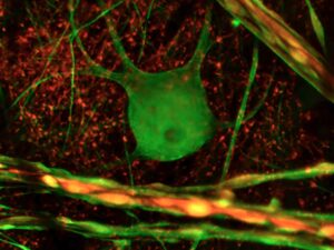
Título: Neurona gigante de retina de Ballena
Aumentos: Microscopio de fluorescencia Zeiss Axio Imager 2, Objetivos Plan Apochromat 20x NA:0,8 d:0,5. Cámara Axiocam MRM Zeiss, Software ZEN Blue
Autores: Elena Vecino Cordero y Luis López Vecino
La fotografía corresponde a una neurona ganglionar de la retina de una ballena (Balaenoptera pysalus), de 18 metros de largo y 20 toneladas.
El ojo del mamífero mas grande del planeta pesaba 1kilo y fue procesado pocas horas después de morir en la playa. La fotografía de fluorescencia muestra la neurona marcada con un anticuerpo contra dos tipos de neurofilamentos HP (verde) y H (rojo). Los neurofilamentos son proteínas fibrosas que tienen una función estructural, formando el citoesqueleto celular. La neurona de la fotografía es una de las encargadas de transmitir el mensaje desde el ojo hasta el cerebro, por lo que, en la ballena, los axones de estas neuronas recorren varios metros a través del nervio óptico antes de llegar a su relevo en el cerebro. En la ballena, varias de estas neuronas miden hasta 100 micrómetros de diámetro celular, mas del doble que en humanos, por lo que se han denominado neuronas gigantes. Algunos axones con forma arrosariada, que aparecen en primer plano de color amarillo, indican que tienen los dos tipos de neurofilamentos, y tienen un diámetro de varios micrómetros, muy superior a lo normal, indicando las largas distancias que tienen que recorrer
2022
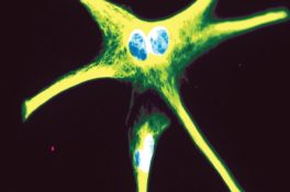 Pinto-Benito, D., Paradela-Leal, C., Ganchala, D., de Castro-Molina, P., & Arevalo, M.-A. (2022). IGF-1 regulates astrocytic phagocytosis and inflammation through the p110α isoform of PI3K in a sex-specific manner. Glia, 70( 6), 1153– 1169. https://doi.org/10.1002/glia.24163
Cover Illustration: Confocal microscopy image of primary culture of female astrocytes, stimulated with bacterial lipopolysaccharide (LPS) and treated with insulin-like growth factor (IGF-1). The image shows the reactive morphology of the cytoskeleton (glial fibrillary acidic protein-green), induced by LPS. IGF-1 cannot reduce reactivity in female astrocytes, only in male astrocytes, indicating that male and female astrocytes show different inflammatory outcomes with LPS administration and IGF-1 treatment. (See Pinto-Benito, D., et al.
2021
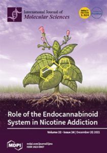 Saravia, R., Ten-Blanco, M., Pereda-Pérez, I., and Berrendero, F.
Int. J. Mol. Sci. 2021, 22(24), 13316
Nicotine, the main psychoactive component in tobacco smoke, plays a major role in tobacco addiction, producing a high morbidity and mortality in the world. A great amount of research has been developed to elucidate the neural pathways and neurotransmitter systems involved in such a complex addictive behavior. The endocannabinoid system, which has been reported to participate in the addictive properties of most prototypical drugs of abuse, is also implicated in nicotine dependence. This review summarizes and updates the main behavioral and biochemical data involving the endocannabinoid system in the rewarding properties of nicotine as well as in nicotine withdrawal and relapse to nicotine-seeking behavior. Promising results from preclinical studies suggest that manipulation of the endocannabinoid system could be a potential therapeutic strategy for treating nicotine addiction
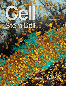
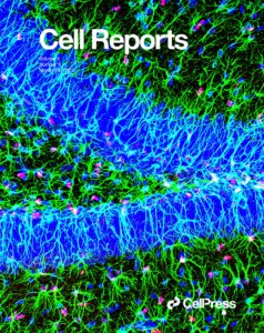 Li L, Medina-Menéndez C, García-Corzo L, Córdoba-Beldad CM, Quiroga AC, Calleja Barca E, Zinchuk V, Muñoz-López S, Rodríguez-Martín P, Ciorraga M, Colmena I, Fernández S, Vicario C, Nicolis SK, Lefebvre V, Mira H, Morales AV. SoxD genes are required for adult neural stem cell activation. Cell Reports, 2022 Feb 1;38(5):110313. doi: 10.1016/j.celrep.2022.110313.
On the Cover: The adult neurogenic niche in the hippocampus is maintained through activation of reversibly quiescent neural stem cells (NSCs). The cover highlights NSCs in the hippocampal dentate gyrus with radial processes GFAP+ (green) expressing transcription factor Sox6 (red). Most of the NSCs are quiescent except for those expressing MCM2 (activated, white). Li et al. report that Sox6 and Sox5 are required for NSC activation and for the generation of new neurons in the adult hippocampus in mice. Image credit: Cristina Medina-Menéndez
2018
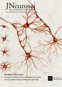 Sanz-Rodriguez, M, Gruart, A.Z, Escudero-Ramírez, J, de Castro, F., Delgado-García, J.M., Wandosell, F and Cubelos, B. R-Ras1 and R-Ras2 Are Essential for Oligodendrocyte Differentiation and Survival for Correct Myelination in the Central Nervous System. J. Neurosci., 38(22), 5209-5219
This watercolor, inspired by the drawings of Ramón y Cajal, shows an oligodendrocyte ensheathing two axons, accompanied by illustrations of the different maturation stages through which the oligodendrocyte progenitor cells pass before becoming mature, myelinating oligodendrocytes. The GTPases R-Ras1 and R-Ras2 are essential regulators of oligodendrocyte development and myelination. Drawing by Daniel Belchi.
2017
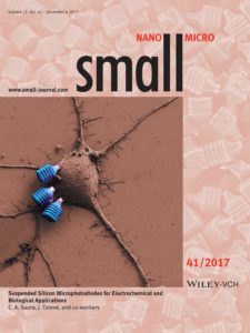 Vargas-Estevez C., Duch, M., Duque, M, del Campo F.J., Enríquez-Barreto, L., Murillo G., Torras N., Plaza Plaza J.A., Saura C.A., Esteve, J. Suspended silicon micro-photodiodes for electrochemical and biological applications (2017). Small. 13(41)
In this article by Carlos A. Saura, Jaume Esteve, and co-workers, show fabrication of suspended silicon microphotodiodes as a simple and cost-effective wire-less microtool for delivering photocurrent in liquid media. Electrical and electrochemical characterization of microphotodiodes under on/off illumination conditions was performed. Their effectiveness as remote photovoltaic cells for life science applications was evaluated in vitro in mouse cultured neurons. The picture shows silicon microphotodiodes bound to the cell surface of a neuron captured by scanning electron microscopy.
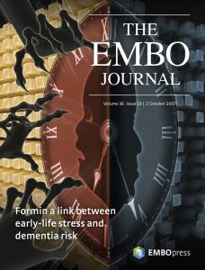 Agís-Balboa RC, Pinheiro PS, Rebola N, Kerimoglu C, Benito E, Gertig M, Bahari-Javan S, Jain G, Burkhardt S, Delalle I, Jatzko A, Dettenhofer M, Zunszain PA, Schmitt A, Falkai P, Pape JC, Binder EB, Mulle C, Fischer A, Sananbenesi F. Formin 2 links neuropsychiatric phenotypes at young age to an increased risk for dementia. EMBO J. 2017 Oct 2;36(19):2815-2828.
Deregulation of the actin cytoskeleton regulator Formin 2 is a common denominator of post-traumatic stress disorders and age-dependent Alzheimer’s disease phenotypes, possibly explaining their clinical connection. Agis-Balboa et al. provide molecular insight to the phenomena that people suffering from PTSD at young age have an increased risk to develop Alzheimer’s disease. The data suggest that the Formin 2 protein and aberrant gene-expression play a key role.
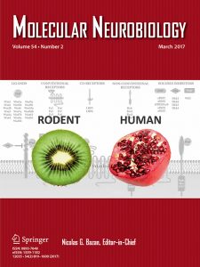 Gonzalez-Fernandez C, Arevalo-Martin A, Paniagua-Torija B, Ferrer I, Rodriguez FJ,Garcia-Ovejero D. Wnts Are Expressed in the Ependymal Region of the Adult Spinal Cord. Mol Neurobiol2016 Oct 8. doi: 10.1007/s12035-016-0132-8.
In rodent spinal cord, neural precursors/stem cells are arranged around the central canal, like the seeds around the central placenta (white core) of the kiwi fruit. But in adult humans central canal is collapsed, and ependymal cells rearrange forming cell aggregations like those formed by seeds in the pomegranate. In the current paper, we show that Wnts are expressed both in rats and human ependymal region, but with a different pattern, suggesting a role for this family of proteins in both species, but probably different functional implications in each of them. Kiwi fruit and pomegranate pictures are under Creative Commons CC0 License and have been obtained from the free repository Pixabay.
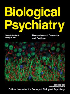 Parra-Damas A., Chen M., Enríquez-Barreto L., Ortega L., Acosta S., Camats Perna J., Fullana N., Aguilera J., Rodríguez-Alvarez J., and Saura C.A. CRTC1 function during memory encoding is disrupted in neurodegeneration. Biol Psychiatry 2017; 81 (2): 111-23.
Landscape of pyramidal neurons showing localization of the transcriptional coactivator CRTC1 (green) in the nucleus (blue) and surrounding MAP2-positive dendrites (red) in the adult mouse hippocampus after memory training. Nuclear translocation and function of CRTC1 is disrupted in a mouse model displaying associative memory deficits and Alzheimer´s disease like-pathological features.
 M. Valero, RG. Averkin, I. Fernandez-Lamo, J. Aguilar, D. Lopez-Pigozzi, JR. Brotons-Mas, E. Cid, G. Tamas, and L. Menendez de la Prida. Mechanisms for selective single-cell reactivation during offline sharp-wave ripples and their distortion by fast ripples. Neuron 2017; 94 (6): 1234-47.
The study explores the synaptic mechanisms of firing selectivity by CA1 pyramidal cells during sharp-wave ripples. Using unsupervised clustering of ripple waveforms, the authors show how an intracellular synaptic depolarization determines firing preference during some sharp-wave ripple events. This mechanism is impaired in epileptic rats and cells fire indiscriminately following global synaptic influences. The image depicts the waves beating the seashore and two spiky shadows at the edge choosing a ripple to jump. Image from Adam Goldberg, used under license from Shutterstock.com.
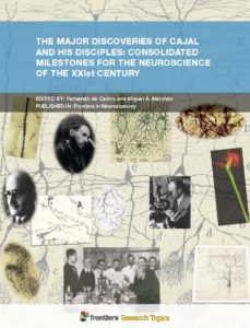 de Castro, F., y Merchán, M. A., editores (2017). “The Major Discoveries of Cajal and His Disciples: Consolidated Milestones for the Neuroscience of the XXIst Century”. Ed. Frontiers Media, Lausana (Suiza). ISBN 978-2-88945-066-4, 163 páginas (doi: 10.3389/978-2-88945-066-4).
Composition images illustrating the articles of the book. Upper row (from left to right): dendritic spines (Golgi method), Santiago Ramón y Cajal, dividing astrocytes (by Santiago Ramón y Cajal), and Golgi staining (by Miguel Marín-Padilla). Middle row: Pío del Río-Hortega, oligodendrocytes, microglia (both drawings by Río-Hortega), Fernando de Castro at the microscope (Wiesbaden, 1950) and current visualization of a neuronal growth cone. Bottom row: innervation of the carotid región (original drawing by Fernando de Castro), Cajal´s lesson to his disciples (including Gonzalo R. Lafora, Gayarre, Sacristán, Francisco Tello and Nicolás Achúcarro), neuron (original drawing by Rafael Lorente de Nó).
2016
 Agís-Balboa RC, Pinheiro PS, Rebola N, Kerimoglu C, Benito E, Gertig M, Bahari-Javan S, Jain G, Burkhardt S, Delalle I, Jatzko A, Dettenhofer M, Zunszain PA, Schmitt A, Falkai P, Pape JC, Binder EB, Mulle C, Fischer A, Sananbenesi F. Formin 2 links neuropsychiatric phenotypes at young age to an increased risk for dementia. EMBO J. 2017 Oct 2;36(19):2815-2828.
Deregulation of the actin cytoskeleton regulator Formin 2 is a common denominator of post-traumatic stress disorders and age-dependent Alzheimer’s disease phenotypes, possibly explaining their clinical connection. Agis-Balboa et al. provide molecular insight to the phenomena that people suffering from PTSD at young age have an increased risk to develop Alzheimer’s disease. The data suggest that the Formin 2 protein and aberrant gene-expression play a key role.
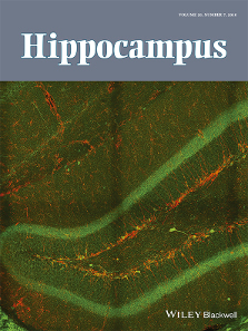 Gradari, S., Pérez-Domper, P., Butler, R.G., Martínez-Cué, C., de Polavieja, G.G. and Trejo, J.L. The relationship between behavior acquisition and persistence abilities: Involvement of adult hippocampal neurogenesis. Hippocampus 2016; 26 (7): 857-74. doi: 10.1002/hipo.22568. doi: 10.1002/hipo.22568.
Doublecortin (DCX)-Calretinin(CLR) immunolabeling of the hippocampal dentate gyrus of a 4 month old male mice (Mus musculus). DCX+(red) and CLR+(green) neurons in a section from a control (no treatment) mouse, where both DCX+/CLR+ and DCX+/CLR+ can be appreciated. This dual labeling lets to count two different subpopulation of immature, differentiating neurons in two successive stages of maturation with one double immunohistochemistry reaction. Cell counts were performed in these type of sections by using the U-disector and confocal microscopy. The cover image, by J.L. Trejo et al., is based on the Research Article The relationship between behavior acquisition and persistence abilities: Involvement of adult hippocampal neurogenesis.
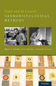 Merchán, M.A., DeFelipe, J., y de Castro, F. editores (2016) “Cajal and De Castro´s Neurohistological Methods”. Oxford University Press, Oxford (Gran Bretaña). ISBN 978-0-19-022159-1, 252 páginas.
Portraits of Santiago Ramón y Cajal and Fernandro de Castro, with a recent picture of a historical tray full of original histological slides generated by the portraited scientists.
 Sierra A, de Castro F, del Río-Hortega J, Iglesias-Rozas J, Garrosa M, y Kettenmann H (2016) The `big-bang´ for modern glial biology: translation and comments on Pío del Río-Hortega 1919 series of papers on microglia. Glia, 64:1801–1840 (doi:10.1002/glia.23046). “Cover page” seleccionada para el Volume 64 (Issue 11, pages 1795–2040).
Pío del Río-Hortega´s original drawings of microglia and oligodendrocytes, cells that he identified unravelling the secrets of the Cajal´s “third element of the nervous system”.
2014
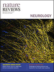 Barcia, C. y Barba, N. Nature Reviews Neurology 2014; 10 (2).
Glial network in the human cortex. Astrocytes with their long processes (yellow) are immunostained with GFAP antibodies, and microglia with their shorter processes are visualized with IBA1 antibodies (blue). Glial cells form an extensive and uniform network along the brain parenchyma that is crucial for the correct functioning of the CNS. Research on glial cells provides important insights into the pathogenesis of many neurodegenerative diseases and neurological disorders.
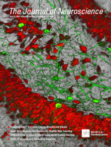 Parra-Damas A., Valero J., Chen M., España J., Martin E., Ferrer I., Rodríguez-Alvarez J. and Saura C.A. CRTC1 activates a transcriptional program deregulated at early Alzheimer´s disease- related pathological stages. J Neurosci 2014; 34 (17): 5776-87.
Confocal immunofluorescence image showing localization of the transcriptional coactivator CRTC1 in neurons of the adult mouse hippocampus after adeno-associated viral-CRTC1 gene therapy. CRTC1 (green) localizes in neurites and nuclei of CA3 hippocampal and dentate gyrus neurons (red, NeuN). Authors: Núria Barba, Arnaldo Parra-Damas and Carlos A. Saura.
2013
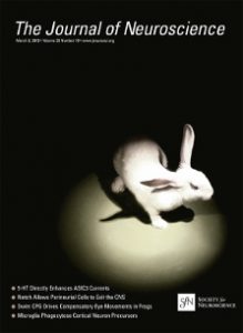 Leal-Campanario, R., Delgado-García, J.M., Gruart, A. Electrical stimulation of the rostral medial prefrontal cortex inhibits reflex blinks and the expression of conditioned eyelid responses, but does not affect its acquisition, in behaving rabbits. Journal of Neuroscience 2013; 33 (10): 4378-86. ISSN: 0270-6474.
The rostromedial prefrontal cortex in rabbits is a potent inhibitor of acquired eyelid responses until the need for the acquired response is fully confirmed. In this regard, the rostromedial prefrontal cortex seems to act as flip-flop mechanism controlling the expression of reflex and acquired behaviors depending on the animal environmental circumstances. Photography by R. Leal-Campanario, J.A. Santos and J.C. López-Ramos; editing by R. Leal-Campanario.
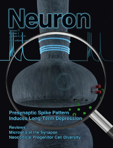 Rodríguez-Moreno, A., González-Rueda, A., Banerjee, A., Upton, A.L., Craig, M.T. and Paulsen, O. Presynaptic self-depression at developing neocortical synapses. Neuron 2013 Jan 9; 77 (1): 35-42. doi: 10.1016/j.neuron.2012.10.035.
Activity-dependent plasticity is important for synaptic refinement during cortical development. The cover shows that a specific spike pattern in individual presynaptic layer 4 neurons (blue traces) can induce long-term depression (LTD) without the involvement of the postsynaptic neuron. This presynaptic spike pattern-dependent LTD (p-LTD) requires release of glutamate (green spheres) activating presynaptic NMDA receptors (red rods).
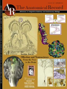 García-González, D., Murcia-Belmonte, V., Clemente, D., y de Castro, F. (2013) Olfactory system and demyelination. Anat. Rec. 296, 1424-1434. “Cover page” seleccionada para el volumen 296, número 9.
Upper-right schema in the composition: schematic representation of our hypothesis that part of the cases of Kallmann síndrome would be explined by deffects in demyelination and would be there linked to leukodystrophies.
2011
 Encinas, J.M., Michurina, T.V., Peunova, N., Park, J.H., Tordo, J., Peterson, D.A., Fishell, G., Koulakov, A. and Enikolopov, G. Division-coupled astrocytic differentiation and age-related depletion of neural stem cells in the adult hippocampus. Cell Stem Cell 2011; 6; 8 (5): 566-79. doi: 10.1016/j.stem.2011.03.010.
Production of new neurons in the adult brain goes down with age and this decline may underlie age-related cognitive impairment. Encinas et al. (pages 566–579) show that aging is associated with the decrease in the number of neural stem cell in the hippocampus. These cells disappear, after a rapid series of divisions, via their conversion into astrocytes. The image depicts stem cells (teal) and neuronal nuclei (yellow), with the autumn trees with falling leaves symbolizing the loss of cells during aging. Image created by Elena Nikanorova, Ann-Shyn Chiang, and Grigori Enikolopov.
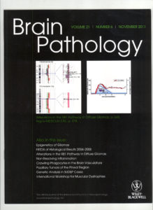 Moliné-Velázquez, V., Cuervo, H., Vila del Sol, V., Ortega, M.C., Clemente*, D., y de Castro*, F. (2011) Myeloid-derived suppressor cells limit the inflammation by promoting T lymphocyte apoptosis in the spinal cord of a murine model of multiple sclerosis. Brain Pathol. 21, 678-691 (doi:10.1111/j.1750-3639.2011.00495.x). «Cover page» seleccionada para el volumen 21
Production of new neurons in the adult brain goes down with age and this decline may underlie age-related cognitive impairment. Encinas et al. (pages 566–579) show that aging is associated with the decrease in the number of neural stem cell in the hippocampus. These cells disappear, after a rapid series of divisions, via their conversion into astrocytes. The image depicts stem cells (teal) and neuronal nuclei (yellow), with the autumn trees with falling leaves symbolizing the loss of cells during aging. Image created by Elena Nikanorova, Ann-Shyn Chiang, and Grigori Enikolopov.
2010
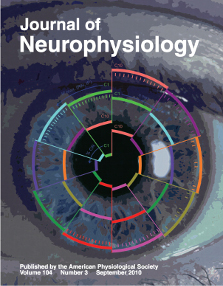 Elena Porras-García*, Raudel Sánchez-Campusano, David Martínez-Vargas, Eduardo Domínguez-del-Toro, Jan Cendelín, Frantisek Vožeh, José M. Delgado-García. Behavioral Characteristics, Associative Learning Capabilities, and Dynamic Association Mapping in an Animal Model of Cerebellar Degeneration. Journal of Neurophysiology 2010; 104: 346-65; doi:10.1152/jn.00180.2010.
Representation of the three main regularities in the instantaneous firing (IF) activities of red nucleus (RN) and cerebellar interpositus (IP) neurons observed during classical eyeblink conditioning. The message is that the learning trace in these structures is dissipated because the level of the dynamic associations (η max., middle spiral) decreases across training (from C1 to C10).
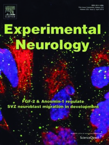 García-González, D., Clemente, D., Coelho, M., Esteban, P., Soussi-Yanicostas, N. y de Castro, F. (2010) Dynamic roles of FGF-2 and Anosmin-1 in the migration of neuronal precursors from the subventricular zone during pre- and postnatal development. Exp. Neurol. 222, 285–295 (doi:10.1016/j.expneurol.2010.01.006). “Cover page” seleccionada para el volumen 222, número 2
Schema in the right: summary of the entry of Myeloid-Derived Supressor Cells (MDSCs) into the CNS parenchyma after EAE immunization and BBB disruption. The model proposes that MDSCs enter to limit inflammation and subsequent demyelinating damage, and then leave the CNS parenchyma, again.
2008
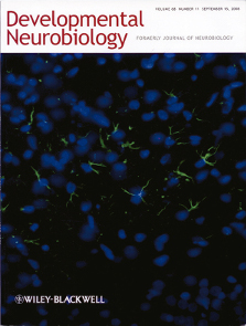 Llorente, R., Llorente-Berzal, A., Petrosino, S., Marco, E.M., Guaza, C., Prada, C., López-Gallardo, M., Di Marzo, V. and Viveros, M.P. (2008) Gender dependent cellular and biochemical effects of maternal deprivation on the hippocampus of neonatal rats; a possible role for the endocannabinoid system. Dev Neurobiol 2008; 68 (11): 1334-47. ISSN 1932-8451.
Microphotograph showing glial fibrillary acidic protein (GFAP) positive cells (green) with cellular nucleus staining with DNA-binding fluorescent dye 4’, 6-diamidino-2-phenylindole dihydrochloride (DAPI, blue) from the stratum radiatum and lacunosum-moleculare of hippocampal CA1 area of a rat.
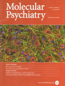 Gascón S, Sobrado M, Roda JM, Rodríguez-Peña A, and Díaz-Guerra M. Excitotoxicity and focal cerebral ischemia induce truncation of the NR2A and NR2B subunits of the NMDA receptor and cleavage of the scaffolding protein PSD-95. Mol Psychiatry 2008;13 (1):99-114.
Double immunohistochemistry of rat cerebral cortex showing partial co-localization of subunits NR2A and NR2B of the NMDA receptor (red) and PSD-95 (green) in the membrane of neurites. The cell nuclei were stained with TO-PRO (blue). In a rat model of brain ischemia, a parallel decrease of PSD-95 and NR2 subunits was observed in the infarcted cortical region.

Hazte socio aquí
Contribuye al progreso de la Neurociencia

Colabora con la Sociedad
¿Quieres publicar alguna noticia o evento?